IPad Apps

Rocks and Minerals

Structure of earth

Simple Machines
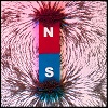
Magnets

Animal Adaptations

Plant Adaptations

Diseases
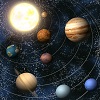
Solar System
Welcome to FunAppSchool
IPad and IPhone Apps
Educational Games for IPad and IPhone. English Grammar and Science Apps for Elementary and Middle School Kids.
Microscopes
Microscopes are optical (eye) instruments that allow us to see small objects enlarged several times over.
The difference between a microscope and telescope is that the telescope makes distant objects appear nearer, while the microscope allows us to see objects that are near, just clearer.
The mechanism that allows microscopes to do their job is called magnification. It is the power to enlarge the image of whatever objects being looked at multiple times.
In simple lenses, if a lens is said to have a magnification of 2X it means that the lens can magnify the image of the object 2 times.
The more complex lenses such as those in compound microscopes, have more complex ways of calculating the total magnification.
Along with magnification comes resolution. Resolution depends on the light rays.
The resolution of a microscope is defined as the shortest distance between two points that a microscope can clearly show as being separate.
Generally, a lens that provides higher magnification also provides better resolution.
Historians believe that Hans and Zacharias Jansen invented the first microscope in 1600, which used two lenses and could increase the size of the object’s picture about thirty times.
In the late 1660s, Antony van Leeuwenhoek used a powerful magnifying glass to examine plant and animal tissues, blood cells, minerals and fossils.
Even though his was not a microscope, he discovered microscopic animals and plaque formations on teeth with his apparatus, which could magnify an object about 200 times.
For the next hundred to two hundred years or so, the design of the microscope did not change.
The lenses however, went through many changes. Purer glass and different shaped lenses were used to see under which conditions the picture was the clearest.
Ernst Abbe made a discovery in the late 1880s, which has influenced microscope lens technology even today.
He found that lenses dipped in a certain type of oil prevented light from playing tricks on the lens, which ruined the picture.
These lenses were also able to achieve the highest magnification power. This meant that the picture was enlarged to over 1000 times. These are still used today.
In the 1931, the electron microscope was invented. This instrument directs a beam of electrons at the specimen being examined.
The electrons are absorbed or scatter about the cell and the pattern they create is captured as an image on the electron-sensitive photo plate.
This great instrument allows scientists to examine small parts that are magnified over a million times.
Unfortunately this machine cannot be used to examine living cells, but they are being improved using digital technology so that even living cells can be examined.
In 1981 Gerd Binnig and Heinrich Rohrer invented a microscope that allows 3-D imaging even at the tiniest microscopic level. This was called the scanning tunnelling microscope.
They won the Nobel Prize for Physics for it in 1986 and it remains the most powerful microscope to date.
Even though there are no microscope manufacturers in America, Germany, China and Japan invent state of the art microscopes.
The most common microscope found in most hospitals, research centres, and even your school laboratory is called a compound microscope.
This instrument operates by using a series of lenses to collect and focus the light transmitted through the specimen being looked at.
IPad/IPhone